Regular prenatal check-ups help to recognise risks early and to accompany your pregnancy safely. On this page you will find an overview of the screening tests and ultrasound examinations we offer in our practice in Limassol – from first-trimester screening and NIPT to Doppler and 3D/4D ultrasound.

First-trimester antenatal pregnancy screening, conducted between the 12th and 14th week, is a non-invasive test to identify potential genetic or structural abnormalities in the fetus. Integral to antenatal care, it enables early detection of anomalies, facilitating prompt and appropriate action.
The first-trimester screening can provide indications of genetic anomalies such as Down syndrome (Trisomy 21) and other chromosomal disorders. Additionally, structural abnormalities of the fetus can be detected through this examination.
First-trimester prenatal pregnancy screening is a valuable test offering early reassurance about your baby’s health. It enables us to identify potential risks, prompting further tests or consultations if necessary. The measurement of hormones bhcG and PAPP-A, coupled with ultrasound, enhances the accuracy of risk assessment.
Non-invasive prenatal testing (NIPT) is a non-invasive prenatal screening test without risk to the pregnancy, providing expectant parents with crucial information about the health of their unborn baby. Unlike traditional invasive tests like amniocentesis or chorionic villus sampling, NIPT carries no risks for either the mother or the fetus.
The NIPT is based on a simple blood draw from the mother. Fetal DNA fragments, known as cell-free fetal DNA, circulate in the maternal blood. By analyzing these DNA fragments, we can gather genetic information about the baby, including the risk of certain chromosomal disorders such as trisomy 21 (Down syndrome), trisomy 18 (Edwards syndrome), and trisomy 13 (Patau syndrome).
I work as a gynecologist in Limassol and I am pleased to welcome you to my practice.
While non-invasive prenatal testing (NIPT) is notably accurate, it’s crucial to underscore that it assesses likelihood rather than a definitive diagnosis. An invasive test becomes necessary for a conclusive diagnosis in the event of an abnormal result.

Preeclampsia is a distinct manifestation of high blood pressure coupled with proteinuria, occurring during pregnancy or post-childbirth. While the exact cause remains incompletely understood, it is hypothesized that issues related to blood flow to the placenta and inflammatory responses are pivotal contributors to its development.
During the first trimester of pregnancy, a secure method exists to calculate the likelihood of developing a particular disease. This allows for timely therapeutic intervention to prevent the disease and identifies pregnant women requiring more intensive monitoring.
A specialized computer program integrates maternal characteristics, blood pressure readings, Doppler examination of uterine vessels, and the values of two blood factors (PAPP-A, PlGF) to compute the probability of the disease.
In instances of high probability, treatment involving daily aspirin is essential, significantly reducing the risk of pre-eclampsia. This is followed by heightened monitoring throughout the pregnancy, substantially diminishing the risk of complications for both the pregnant woman and the fetus.
Anticipating and screening for the risk of pre-eclampsia is a vital component of our all-encompassing care, ensuring early diagnosis and effective management of potential complications.
Should you have questions, concerns, or a desire to delve deeper into this screening process, we are here to assist you. Your well-being and that of your baby remain our utmost priority.
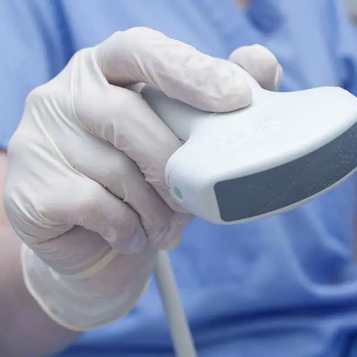
Doppler ultrasound in pregnancy is a non-invasive examination providing crucial insights into your baby’s development and well-being. By assessing the speed of blood flow in the fetus and uterine blood vessels, we draw conclusions about the oxygen and nutrient supply to the baby. Maintaining balanced blood flow is essential for ensuring your baby’s healthy growth and development.
During this examination, particular ultrasound waves are used to measure the blood flow velocity in the baby’s blood vessels and the uterus (maternal placenta). We can infer the oxygen and nutrient supply to the baby based on the determined blood flow values. Balanced blood circulation is essential for your child’s healthy growth and development.
This examination is not only gentle and painless but also highly informative. Through regular use of fetomaternal Doppler, potential issues can be detected early, and appropriate measures can be taken to protect your baby’s health and your well-being.
At our gynecological practice in Limassol, we attach the utmost importance to carefully monitoring your pregnancy. Embryometric Doppler is essential to our comprehensive care and is always individually tailored to your needs. If you have any further questions, please do not hesitate to contact us. As an experienced gynecologist in Limassol, I look forward to welcoming you to my office.

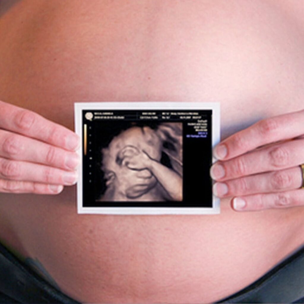
4D ultrasound with “Realistic View” is an exciting and innovative technology that allows us to offer you and your partner a realistic image of your unborn baby. This advanced method can produce amazingly detailed images and even fetal movement sequences.
Conventional 2D Ultrasound shows you black-and-white images of your baby in a two-dimensional representation. In contrast, 3D Ultrasound creates lifelike three-dimensional images that add depth and contour to your baby. With 4D Ultrasound, we can create real-time motion recordings that capture moments like kicking or touching the face.
This innovative technology’s “Realistic View” function is at the heart. It enables us to display the skin and surface of your unborn baby even more realistically. As a result, you see amazingly vivid images of your baby and can admire, for example, the tiny fingernails, facial features, and body details.
3D/4D Ultrasound with “Realistic View” is part of our efforts to provide you with a unique and unforgettable experience. We are here to assist you if you have questions or are interested in this procedure.

Screening for infectious diseases such as rubella, toxoplasmosis, cytomegalovirus (CMV), parvovirus B19 (fifth disease), and chickenpox is a significant step in identifying potential infection risks during pregnancy. Certain infectious diseases can harm the unborn baby and lead to complications. By detecting and treating these infections early, we can ensure the best possible care for you and your baby.
A blood sample is collected to screen for antibodies against specific infectious diseases. This test helps identify pregnant women who lack immunity and may face an elevated risk of infection upon exposure to pathogens.
Upon obtaining the results, couples are informed and provided with appropriate guidance based on the analysis.

Gestational diabetes, a diabetes variant occurring during pregnancy, presents potential risks for both the mother and baby. The blood glucose loading test, also called the glucose tolerance test or oral glucose tolerance test (oGTT), is a crucial diagnostic tool for early identification of gestational diabetes. This test involves monitoring blood sugar levels after consuming a specialized sugar mixture.
The blood glucose loading test is typically conducted between the 24th and 28th week of pregnancy. Undertaking the test during this specific timeframe is crucial, allowing for an early diagnosis and providing ample time to implement measures that minimize risks for both the mother and fetus.
An abnormal test result may suggest challenges in regulating blood sugar during pregnancy, indicating a positive test for gestational diabetes.
It’s essential not to be overly concerned if your test shows irregular values. A positive result signals the need for additional examination and specialized treatment from us, ensuring your and your baby’s well-being.
Dr. Roukoudis and his team will be glad to answer your questions and plan the next steps with you.